Catalog
-
Catalog
- Agriculture
- Apparel
- Automobiles & Motorcycles
- Beauty & Personal Care
- Business Services
- Chemicals
- Construction & Real Estate
- Consumer Electronics
- Electrical Equipment & Supplies
- Electronic Components & Supplies
- Energy
- Environment
- Excess Inventory
- Fashion Accessories
- Food & Beverage
- Furniture
- Gifts & Crafts
- Hardware
- Health & Medical
- Home & Garden
- Home Appliances
- Lights & Lighting
- Luggage, Bags & Cases
- Machinery, Hardware & Tools
- Measurement & Analysis Instruments
- Mechanical Parts & Fabrication Services
- Minerals & Metallurgy
- Office & School Supplies
- Packaging & Printing
- Rubber & Plastics
- Security & Protection
- Service Equipment
- Shoes & Accessories
- Sports & Entertainment
- Telecommunications
- Textiles & Leather Products
- Timepieces, Jewelry, Eyewear
- Tools
- Toys & Hobbies
- Transportation
Filters
Search
joints and ligaments specimen,medical equipment
Beijing, China
86-10-63977620

Guoxin Chen
Contact person
Basic Information
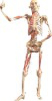
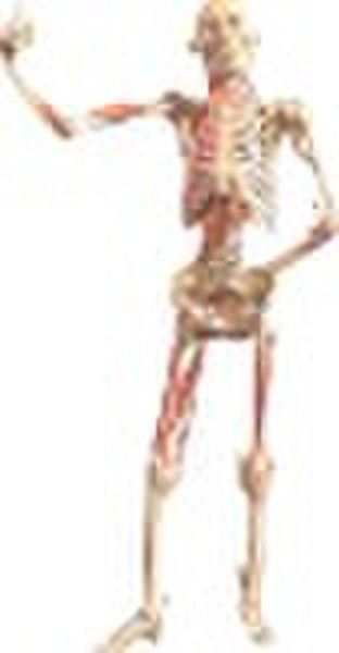
Synopsis: Due to the limit of time and anatomical skills, the delicate dissection of all the joints and ligaments is unavailable in anatomical teaching class. Therefore, the teaching specimens are necessary for the students to understand and remember. This specimen displays the skeleton, joints and ligaments. Around the joints, some deep muscles are remained so that the origins are clear, which will be helpful for the students to understand the role of the muscles during movement. Displaying contents: The joints of skull:1. Sutures : lambdoid suture, coronal suture and sagittal suture2. Temporomandibular joint: The left articular capsule is integrated and the lateral ligament is remained. The right articular capsule is sagittally cut to display the articular disc and cavity. The joints of the trunk bones:1. The joints of vertebrae: anterior longitudinal ligament, yellow ligament, interspinal ligament, superaspinal ligament (ligamentum nuchae), intertransverse ligament, anterior and posterior atlatooccipital membranes. One vertebral body is partly removed to display intervertebral disc (mucleus pulposus and annulus fibrosus). 2. Thoracic joints: The sternum is cut coronally to display the sternocostal joints and sternoclavicular joint (articular disc) on right side. On the other side, intercostals externi, intercostals interni, levator scapulae, longus capitis, longus colli, scalenus anterior, medius and posterior are displayed.The joints of the bones of limbs 1. The joints of the upper limbs:2. The joints of the lower limbs:Pelvic ligaments: iliolumbar ligament, dorsal and ventral sacroilia ligaments, pectineous ligament, sacrotuberous ligament, sacrospinous ligament and pubic symphysis.
Delivery terms and packaging
Packaging Detail: according to customers' requirement Delivery Detail: as soon as possible
Port: Tianjin xingang
Payment term
Telegraphic transfer
Western Union
-
Payment Methods
We accept:


